Advanced Cervical Spine Treatment by Dr. Ankit Daware – Consultant Spine Surgeon
Understanding the C1–C2 Region
What Is C1–C2 Instability?
Common Causes of C1–C2 Instability
Several conditions may lead to instability at the upper cervical segment. Some of the most common include:
1. Trauma or Injury
High-velocity injuries such as
Road traffic accidents
Sports injuries
Falls from height
can cause fractures, ligament tears, or misalignment.
2. Rheumatoid Arthritis (RA)
RA can weaken ligaments and joints, leading to progressive instability.
3. Congenital Disorders
Conditions such as:
Os odontoideum
Down syndrome
Skeletal dysplasias
may lead to lifelong cervical instability.
4. Degenerative Spine Disease
Wear and tear due to aging can cause ligament laxity and bone changes.
5. Infections or Tumors
Occasionally, infections like tuberculosis or tumors around the cervical spine may damage bone and soft tissue.
6. Inflammatory Disorders
Chronic inflammation weakens supporting structures, causing excessive motion at C1–C2.
Symptoms of C1–C2 Instability
Symptoms may vary depending on severity, but common signs include:
Severe upper neck pain
Headache at the base of the skull
Restricted head rotation
Clicking or grinding sensation
Tingling or numbness in arms
Weakness in limbs
Difficulty walking or balancing
Dizziness or vertigo
In severe cases, breathing or swallowing issues
Although symptoms may start mildly, instability can progress rapidly. Therefore, early evaluation by a spine specialist like Dr. Ankit Daware is essential.
When Is C1–C2 Fusion Needed?
C1–C2 fusion is recommended when instability threatens spinal cord safety or causes chronic pain that does not improve with conservative treatment.
Indications for Surgery:
Persistent neck pain despite physiotherapy or medications
Progressive neurological symptoms
Spinal cord compression on MRI
Unstable fractures
Atlantoaxial subluxation
Rheumatoid arthritis with instability
Tumor or infection-related instability
Congenital instability causing severe symptoms
Early surgical intervention prevents permanent neurological damage and restores spinal stability.
What Is C1–C2 Fusion?
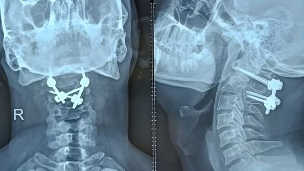
C1–C2 Fusion, also known as Atlantoaxial Fusion, is a procedure performed to stabilize the joint between the atlas and axis. In this surgery, screws and rods or other implants are used to hold the two vertebrae in a fixed position until bones naturally fuse over time.
The goal is to:
Prevent abnormal motion
Protect the spinal cord
Reduce pain
Improve stability
Prevent deformity progression
Types of C1–C2 Fusion Techniques
Dr. Ankit Daware uses the most advanced and safe surgical methods based on each patient’s anatomy and condition.
1. Goel–Harms Technique
The most widely used modern technique, involving C1 lateral mass screws and C2 pedicle/pars screws connected with rods.
Benefits:
Strong fixation
High fusion rate
Safe for most patients
2. C1–C2 Transarticular Screw Fixation
Screws are placed across the C1–C2 joint.
Benefits:
Excellent biomechanical stability
Suitable for selected patients
3. Occipitocervical Fusion (If Needed)
If instability extends upward to the skull base, fusion may include the occiput (back of the skull).
4. Posterior Wiring Techniques
Less commonly used today, but beneficial in selected anatomical situations.
Pre-Surgery Evaluation by Dr. Ankit Daware
Before surgery, a detailed evaluation ensures precise diagnosis and safe surgical planning.
Diagnostic Tests Include:
MRI Scan: evaluates spinal cord compression
CT Scan: studies bone anatomy
Dynamic X-rays: detects abnormal movement
Neurological Examination
Blood Investigations
Dr. Daware explains the condition, discusses options, and ensures complete patient understanding.
How C1–C2 Fusion Surgery Is Performed
The surgery is performed under general anesthesia. Here is a simplified step-by-step process:
Step 1: Positioning
The patient is positioned prone (face down) to allow surgical access.
Step 2: Incision
A small incision is made at the back of the upper neck.
Step 3: Exposure of C1 and C2
Muscles are gently retracted to expose the bone.
Step 4: Screw Placement
Using navigation, fluoroscopy, or 3D imaging:
Screws are inserted into C1 lateral mass
Screws are inserted into C2 pedicle or pars
Step 5: Rod Placement
Rods connect the screws, stabilizing the segment.
Step 6: Bone Grafting
Bone grafts stimulate spinal fusion.
Step 7: Closure
The incision is carefully closed.
With advanced technology and high surgical expertise, the procedure is safe, precise, and effective.
Advantages of C1–C2 Fusion
Patients undergoing surgery with Dr. Ankit Daware benefit from:
Permanent stabilization of the cervical spine
Elimination of abnormal motion
Protection of spinal cord
Reduction in neck pain
Prevention of neurological deterioration
Restoration of balance and posture
Improved quality of life
Recovery After C1–C2 Fusion
Recovery depends on the patient’s condition and the complexity of the procedure.
Hospital Stay
Usually 3–5 days.
Neck Collar
A cervical collar may be advised for a few weeks.
Pain Management
Medications and muscle relaxants help reduce discomfort.
Physiotherapy
Rehabilitation begins gradually to improve posture, strength, and mobility.
Return to Normal Activities
Light activities: 2–4 weeks
Work: 4–8 weeks (depending on job type)
Complete fusion: 3–6 months